Memoir - Edgar F. Meyer
Memoir | Publications | Curriculum Vitae | Videos | Slides | Articles | Obituary
A Crystallographic 4-Simplex
Edgar Meyer
2014
|
In 2003, when Edgar Meyer retired from the Department of Biochemistry & Biophysics at Texas A & M, where he had been a crystallographer for over 36 years, he moved on to the next phase of his career, as a sculptor. He had been a pioneer in 3D computer graphics and database searching, both of which were crucial for the early success of the PDB. Edgar then devoted his energies to the combination of science and art, to his family, and to his vegetable garden.
|
A simplex is the simplest object that can be drawn in a given space domain; "a k-simplex is a k-dimensional polytope - or - the smallest convex set containing the given vertices." For example: the dot, line, and triangle are simplices in 0, 1, and 2D space.
Several threads anchor and define my life: near-death from a childhood accident, fascination with the beauty of crystals, my wife Catarina and our family, and a pervasive urge to create. At age six, I had a life-threatening burn accident with three months in a hospital; survival stamped me with a spiritual sense of purpose. In middle school I remember really enjoying wood shop. In high school, geometry, chemistry, and world history made me excited about learning.
As a teaching assistant in chemistry at North Texas State my senior year, I had the chance to show a movie about crystals and crystal growth. Some in the class may not have appreciated the movie's reference to God as the source of cosmic order, with crystals as a prime example, but crystalline order and beauty was life changing for me - I had to become a crystallographer.
In graduate school at the University of Texas (Austin), I was given a project by Stanley Simonsen. His only crystal of an organometallic complex (bis(3-hydroxyl-1,3-diphenyltriazine) palladium(II) ) was mounted, and I set about measuring diffraction intensities from Weissenberg photographs with a densitometer. After accidentally knocking off the crystal, I spent two years learning crystal growth so that I could complete the project; this skill was useful throughout my career. I learned Fortran and assembly language programming and was exposed to scientific computing on the IBM 650 and CDC 1604.
At my first ACA meeting in Boulder (1961) I met and had a beer with Peter Pauling, who referred me to Jack Dunitz for a post-doc. My two years at the ETH in Zürich with Jack were crucial: 1) the study of the corrin (porphyrin-like) tetrapyrrolic macrocycle of vitamin B12 being synthesized by Eschenmoser and his group - I continued studying porphyrins and polypyrollic structures for the next 10 years; and 2) Catarina Pestalozzi - we have been married for 49 years - Catarina's charm and vitality and wisdom are unchanged with time, even when challenged by my occasional unpredictability. We are enormously proud of our three children and their families.
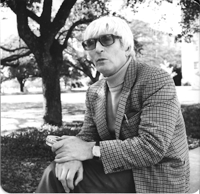
Jack Dunitz at Texas A&M University.
After a year at MIT trying to solve a triclinic structure with mis-indexed upper-layer Weissenberg data I measured by eye, I jumped at the chance to join Cy Levinthal's group, working with the first interactive, real-time computer graphics system available (MIT's Project MAC). I had the chance to write my first graphics program to draw and manipulate molecular structures in 3D.
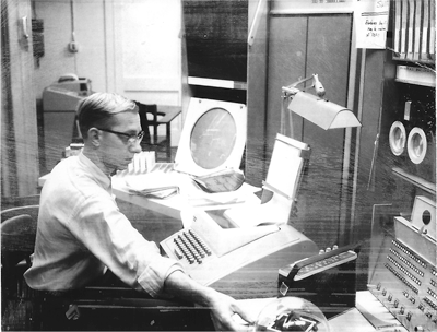
KLUGE display at Project MAC (MIT).
In 1967, with a growing family, I accepted an offer to be an assistant professor at Texas A&M University (TAMU), College Station, Texas, setting up a laboratory studying metalloporphyrin structures. I met Walter Hamilton at the 1968 Tucson ACA meeting, and on short notice Walter arranged for me and my family to spend the summer of 1968 at Brookhaven National Laboratory so that I could use the new Brookhaven RAster Display (BRAD). Starting with crystallographic coordinates, my program DISPLAY drove a color television monitor to draw red/green 3D images of up to 256 atoms, which was crucial for the founding of the Protein Data Bank (PDB) at Brookhaven in 1971. Walter was very supportive, and these summers (1968-1974) at Brookhaven were enormously productive. So, the 3D interactive display of molecular structures was the first apex of the crystallographic simplex and connected immediately with the second apex, the nascent PDB.
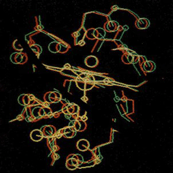
A scoop around the Fe atom in myoglobin.
Teaching duties at TAMU caused me to miss the crucial 1971 Cold Spring Harbor Protein meeting. As things turned out, I could spend only the last six weeks of that summer at Brookhaven to finish program SEARCH, which offered various non-textual search and retrieval tools for studying complex protein structures. With SEARCH and the PDB linked, this was the most productive year of my scientific career. While virtually all search procedures up to this time were text-based, SEARCH could query a structure file for atomic properties. I had at my fingertips, for the first time, a search engine that completed the triangular simplex of structural biology: a database, a search program, and a display to visualize the results in 3D. Walter Hamilton was busy that summer, and so I could not demonstrate the program at Brookhaven. Rather, when he visited me in Texas at the beginning of September, I could run the program remotely at Brookhaven and thus demonstrate the first use of networking in the life sciences (and probably also in chemistry) by remotely selecting a structure (myoglobin) from the PDB database at Brookhaven, defining a search atom (Fe), and extracting all atoms within a chosen radius.
Walter and I obtained a NSF grant to fund a networking project, CRYSNET, linking Brookhaven with TAMU and Fox Chase Cancer Center. While the prevailing graphics technology at the time was monochromatic (black & white), the BRAD system showed the power of color raster graphics directly on the screen. Around this time we began a collaboration with Bob Sparks at Syntex Analytical in Palo Alto.
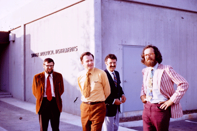
Syntax Analytical (Palo Alto, CA) 1971. L-R: Bob Sparks, Tom Workman, Neville Crooke, Walter Hamilton.
Simultaneously, with the lads in the lab, especially Dave Cullen and Carl Morimoto, we were studying metalloporphyrin structures. The last and one of the most unusual structures was a porphyrin-phosphorus(V) structure studied by Stefano Mangani (now at the Universities of Siena and Florence).
Up to this time the majority of macromolecular structures were solved by building a mechanical (Kendrew=brass) model to fit hand-drawn electron density maps in a device called a Richards Box. In 1975, a graduate student, Marge Legg, working in the laboratory of Al Cotton at TAMU, built the first model of a new protein, staph. nuclease, with interactive 3D graphics, using the visual and manipulative tools of program FIT, begun in my lab by Carl Morimoto and brought to completion by Stan Swanson. Next, in September of 1976, Jim Hogle, a graduate student of M. Sundaralingam's (University of Wisconsin) built the models of both molecules in the asymmetric unit cell of monoclinic lysozyme. A full year passed before another graphics system accomplished a similar feat.
The tools and facilities offered by program FIT thus extended the crystallographic simplex to the next dimension: the tetrahedron. It is easy to overlook the limitation that up to this time the maximum addressable computer memory available was 32 thousand words (vs. 16 gigabytes on my iMac).
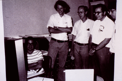
Program FIT Demo: David Klunk, Tom Koetzel, Edgar Mayer, Herb Bernstein, Tom Willoughby.
The issue of Science in 1975 that describes program FIT shows on the cover a stereo tripeptide backbone rotating in space to sculpt out an ethereal image: the first of my virtual sculptures.
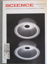
3D image of a tripeptide back-bone rotating in space.
Thanks to an EMBO grant and the hospitality of Robert Huber at Max Planck Institute for Biochemistry (MPIB - Martinsried, Germany), Wolfram Bode of MPIB introduced me to protein chemistry and crystallization. A collaboration with Jim Powers (Georgia Tech) and Jay Fox (Virginia) led to the study of proteolytic enzymes. I was able to spend 10 consecutive summers in Martinsried studying proteolytic enzyme complexes. Short of funds, my lab in Texas had no data collection capability, so I collected enough data each summer to keep the lab busy for the coming year.
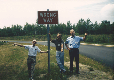
1998 Gordon Research Conference: Edgar Meyer, Xavier Gomis-Ruth, Wolfram Bode.
Computationally, in the late 1980s, the NSF gently urged me to explore molecular dynamics, and I was fortunate to be joined by Bogdan Lesyng and Maciek Geller (University of Warsaw). They, and a graduate student, Gail Carlson (University of Illinois) with the help of Stan Swanson, struggled successfully with supercomputer resources just emerging at that time. Molecular dynamics videos were an eye-opener, but do not qualify as an apex of the simplex for us.
Istvan Botos (NIH), Dachuan Zhang (National Library of Medicine) and my son, Erik (GlaxoSmithKline) were studying metalloproteins from rattlesnake venom (with Jay Fox and Wolfram Bode) and branched out to the study of fire ant chymotrypsin (with funding from the State of Texas). The final structures studied in my lab by Shahram Khademi were a fungal cellulase and the first insect cellulase - of course, from termites, even though the textbooks said they were not supposed to make their own cellulase.
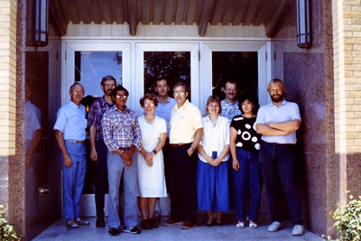
Lab photo: L-R Edgar Meyer, Stan Swanson, Radhakrishnan Rathnachalam (India), Rosie Swanson, Bogdan Lesyng (PL), Dick Rosenfeld, Gail Carlsen, Andi Karrer (CH), Lori Takahashi, Harly Hansasen (DK).
Over these 36 years at Texas A&M, two things kept my lab alive: 1) a sustaining grant from the Robert A Welch Foundation, and 2) a steady flow of bright, energetic students, post docs, and some visiting faculty. La bella Catarina and our children provided stability at home, and for 30+ years Stan Swanson (plus his wife, Rosie) were the brains of the lab when difficult problems came along. Our pursuit of computational and structural biochemistry did not always resonate with the goals of my college (agribusiness), but the internal momentum of the lab made this a pleasant place to work, dream, and create. I taught biochemistry, graduate crystallography, and an honors course, 'Science in Literature,' for many years. I used 3D graphics to make biochemistry teaching more vibrant.
As Catarina and I contemplated the prospects of retirement, we considered moving to Taos, New Mexico. I had always been interested in the art-crystallography connection. So I wrote program SCULPT to generate gcode to control a CNC milling machine. A NSF grant made it possible to install an aged CNC machine donated by Los Alamos National Lab and, after setting up a wood-working shop in Taos, to install a modern machine together with necessary woodworking tools.
Molecular models have been around since 1866, but to have the opportunity to carve a noble hardwood to depict a molecular structure was, and is, a unique blessing. I carved sculptures of amino acids, nucleic acids, and specific compounds as memorials and tributes, some of them privately commissioned. The simplex reaches into the fifth dimension with the addition of molecular sculptures to the apices described above, but the art market has been slow to catch on.
Then, the Smithsonian (National Museum of American History) commissioned a precisely scaled bronze sculpture of the polio virus capsid and the capsid+receptor complex (Jim Hogle lab, Harvard Medical School) as part of the sesquicentennial celebration (2005) of the Salk polio vaccine: www.molecular-sculpture.com/Castings/Polio-bronze/Polio-bronze.html.
A firm in California made the plastic maquettes, and I worked with the Shidoni Foundry in Santa Fe to make the wax models, molds, and bronze castings for the exhibit.
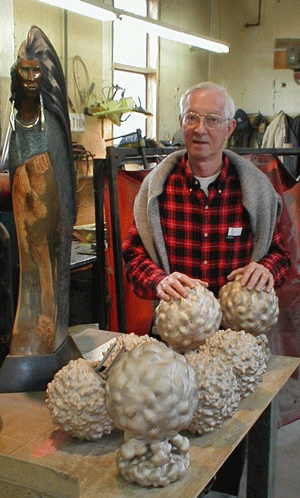
Polio virus sculptures - Shidoni Foundry.
Does 'everyman' know what a vital nutrient or life-saving drug actually looks like (appropriately scaled of course)? And what will future generations know of this "golden age" of crystallography, which successfully elucidated the structures of the major building blocks of life? My answer: we need to place large (2m or taller) precisely scaled bronze or stainless steel sculptures of monumental molecules in public spaces - penicillin in the lobby of a hospital, a block-buster drug at a pharmaceutical laboratory, a carbohydrate, nucleic- or amino-acid model before an athletic field or stadium.
What would such a sculpture look like? And how would it fit in a public space? The same software I use to generate files for rapid prototyping and sculpting also generates high-resolution video files that I have used to make movies of virtual sculptures www.molecular-sculpture.com/WebSampler/Sampler.html.
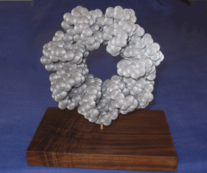
3D sculpture of co-chaperonin (1G31. pdb).
Imagine placing such a sculpture of your structure in front of your building. Imagine children playing on it. Imagine someone asking, "What is it?" = a rare teaching moment.
So, our simplex has five apices, each depicting an adventure in a new direction. Each modest but collectively useful. Nothing original - any of you could have done it - but it was my calling in life and my joy to have the privilege - and honor - to have been associated with gifted, dedicated colleagues and to have contributed in a small way to our discipline as it matured.
Edgar Meyer
Editor's Note: All photos are by the author except where noted otherwise.
|


