- History Home
- People, Leadership & Service
- A Legacy of Excellence
- History & Impact
- Meetings Through the Years
- Resources
Memoir - Jenny Pickworth GluskerMemoir | Publications | Curriculum Vitae | Videos | Slides | Interviews | Articles | Awards | Obituary
As a child I lived in England in the industrial city of Birmingham. Both of my parents were medical doctors and encouraged me in my studies. My father, Frederick Alfred Pickworth, the son of a general practitioner, had been apprenticed to a chemist when 14 years old, and then obtained a degree in chemistry. After working a few years for a chemical company (Burroughs and Wellcome), he went on to medical school at Charing Cross Hospital in London. He then took a research position in Birmingham, where he was primarily interested in brain function, including its response to infection, particularly of the teeth and sinuses (in the days before antibiotics). Nowadays dentists are careful about this problem. He wrote several books on these subjects. My mother, Jane, who was from Scotland, was a medical colleague of his. She had many doctors in her ancestry, including one who went to Russia and was chief physician to three tsars and is mentioned in Tolstoy's "War and Peace" (although as an Englishman, not the Scotsman he really was). My mother had wanted to study foreign languages, but she was in high school in the middle of World War I and there was, by then, a great shortage of young men available to go to medical school because so many of them were already at war. So, it was decided to send girls in high school with good grades to medical school. My mother became a medical student at Glasgow University in 1916. The university had been granting medical degrees to women since the 1890s, but was not used to such large numbers of women students in their medical school classes — about half of her graduating class in 1920 was female. She told me that the professors had to change the mnemonics so that they would be more suitable for such mixed classes. By the time that she graduated the war was over, and she went to Dublin, Ireland, and worked there during the social unrest there in the early 1920s. Finally she accepted a medical post in Birmingham, England, where she met and, after a few years, married my father. From then on she stayed at home to raise her children — my sister who studied history (not a science, said my father), a brother who became a general practitioner and me. Although she was not allowed by the medical community to compete with other doctors in the area, my mother would substitute for local doctors if they had to be away from their practice for any reason. She was very frustrated that she could not continue with a career that she had put so much energy into. For this reason I decided that I would find a way to continue working, even if I had children. So, in my background, many of my parents' friends were women doctors and it was assumed that I would become one.
But I had other ideas. The school that I went to, a public school in the American sense of the word, was newly built, and had excellent scientific laboratories and a chemistry teacher (Dr. Yvonne Way) who inspired me. She had a Ph.D., but decided that she wanted to teach rather than do research, and it was a joy to go to her classes. I still write to her; she is in her 90s. My interest in chemistry had started when I found a book on incompatible medications among my mother's medical textbooks. It explained the chemical processes that resulted when two pills interact unfavorably for the patient. I then acquired a chemistry set that was stored under my bed and was able to mix chemicals and make wonderfully colored solutions and evil-smelling products. Thank goodness I survived that hobby. However, it made me sure that I wanted to be a chemist.
Chance then played a role in my life. Every day, before classes, the entire school of about 700 students (girls only, in those days high schools were generally not coeducational) met in a large hall. One day, in 1947, the principal announced that she had finally received her full Ph.D. degree from Cambridge University. She explained that, until that date, women students who passed all the requirements for an academic degree did not receive the full recognition that men did. Now the University would finally accept them as members of the University with degrees, as for men. The principal wanted us to celebrate with her. I, however, was puzzled. Why should it matter whether you were a woman or a man if you did the same work? I looked up the situation for Oxford University and found they gave degrees and full university membership to women from 1920 on, so I announced I would try to go to Oxford. But my high school teachers all said that they sent science students to Cambridge and did not know too much about chemistry at Oxford. In addition my parents stepped in and said I should go to medical school (which I could do directly from high school). To deal with these problems I made a pact with my father that if I could get into Oxford University that year I would study chemistry. If not I would go to medical school (and not complain).
I did get accepted at the medical school in Birmingham University, but, mindful of my aims, I set off for Oxford to take the entrance exams at Somerville College, the most academic of the women's colleges in Oxford University. It was necessary to take a practical chemistry examination in a laboratory, and the test was to identify an unknown material (in the days of hydrogen sulfide which invariably caused a splitting headache). This "unknown material" consisted of beautiful crystals, but, because I had spent many hours helping my father, an amateur photographer, in his dark room, I knew immediately what they were. They were crystals of sodium thiosulfate or "hypo," used in fixing solutions in photography. During this practical entrance examination every chemical I added to the "unknown material" caused sulfur to precipitate. What a mess! The person at Somerville who supervised me during the exam and then interviewed me for college was Dorothy Hodgkin, a crystallographer and future Nobel Prize winner. During my interview with her I explained about the pact with my father. Well, I was accepted by Oxford University. My father, whom I loved and admired and argued with, never complained that I went there, although, when I had finished college he said he thought I would eventually see the light and go to medical school. Many years later I asked Dorothy Hodgkin if I got accepted to Oxford because I correctly guessed the identity of the unknown material. She said that was not the case, but that she had noted that I knew how to deal with the sulfur that precipitated every time any new chemical was added.
The chemistry course at Oxford took three years, and if one wanted a classified degree, one did an additional year's research. I had a marvelous time. Dorothy Hodgkin was my tutor and we spent many hours in one-on-one weekly tutorials. She was an ideal role model with three children and a supportive husband who was at Balliol College. Her home was a haven for many visiting scientists and it was always interesting to visit her there. For my tutorials Dorothy would insist that I read and comment on all the original papers relevant to the subject she had chosen for my essay that week, so I spent much time in the Radcliffe Science Library. The laboratory experiences were good, but some of the buildings seemed antique compared with those that we had at high school. The design for the 100-year-old octagonal inorganic chemistry laboratory (later replaced) came from the Abbot's Kitchen in Glastonbury Abbey and it had a high ceiling with wooden rafters. But we had some marvelous lecturers Sir Robert Robinson (organic chemistry) and Sir Cyril Hinshelwood (physical chemistry). For my research year I chose to work with Harold W. Thompson, an infrared spectroscopist who was interested in details of molecular structure and had access to some excellent new detection devices so that high-resolution studies could be done. Under his tutelage I determined the interatomic distance in deuterium chloride, for comparison with that of hydrogen chloride being studied by another member of the laboratory. Then I worked on some more complicated molecules, methyl halides, but could only measure moments of inertia and not distances between atoms. However I also met my future husband, Donald Glusker, a Rhodes scholar from the University of California, Berkeley, who was working in the same lab on the spectroscopy of charge-transfer complexes, with Robert Mulliken (on sabbatical from the University of Chicago) as a second advisor.
High-resolution infrared spectroscopy was not, at that time, giving me the information that I wanted. So I started my graduate work in the laboratory of Dorothy Hodgkin, with the aim of learning how to determine molecular structure by X-ray diffraction. The idea was that I would then correlate the structural results I obtained with those from analyses done by use of plane-polarized infrared radiation. The second part of this plan was never carried out. Dorothy was already well-known for her structural work on cholesterol while working with J. D. Bernal. She was the first to show that it is possible for protein crystals to give good diffraction patterns, and, particularly, she determined the chemical formula of penicillin by X-ray diffraction studies in the days when structure determination was very difficult. Dorothy's laboratory and workrooms were in the Oxford University Museum of Natural History that had been built with the help of the artist John Ruskin. We had desks in the very room in which, as commemorated by a plaque on the walls, Thomas Henry Huxley defended Darwin's recently published theory of evolution by natural selection against the Right Reverend Samuel Wilberforce, the then Bishop of Oxford. This took place at a meeting of the British Association in 1860. Dorothy's lab was a busy one and she shared it with Herbert ("Tiny") Powell who was famous for his work on clathrate structures. Jack Dunitz, who, in 1948, had determined the crystal structure of a calciferol derivative, at that time the most complex structure determined by X-ray crystallography, was there when I first started research. Jack was working in an elevated area in the lab that contained the microscopes for crystal viewing, but he was about to go to Caltech. David Sayre had recently left the lab, but everyone was excited about his squaring method for solving structures. Most of the other people in the lab were working on the structure of insulin or vitamin B12. Work on this vitamin, the antipernicious anemia factor, and some of its derivatives, had been in progress in Dorothy's lab for a few years, particularly by John H. Robertson, June Broomhead (later Lindsey), Maureen Mackay and Clara Brink (later Shoemaker). John White at Princeton University was working independently on the same structure, and he collaborated with Dorothy on this throughout the years. Dorothy had decided at that time that no one should work on the much larger insulin molecule for a graduate degree because there was no guarantee that good results could be obtained for it at that time.
Shortly after I had started graduate work some deep red crystals arrived in the lab from Cambridge University. They were of a degradation product of vitamin B12 for which the 5,6-benzimidazole, D-ribofuranoside, 1-amino-2-propanol, phosphate and cyanide groups of the vitamin had been removed and the amide groups had been converted to carboxylic acid groups. The central part of the molecule, containing a cobalt atom, was still present, and that was the part of the chemical formula that was unknown and that we needed to find. Jack Cannon, an Australian, had been trying for some time to crystallize this degradation product that he had made, and finally, in despair, threw into its solution every liquid organic material he had access to, and went to Europe for a holiday. When he came back there were beautiful crystals, never grown again. So I started to work on them. The molecule was large and spectroscopic studies had shown that, like the vitamin itself, its degradation product contained a ring structure similar to (but not the same as) that found in porphyrins. Both the vitamin and Cannon's degradation product contained cobalt atoms which were useful as heavy atoms. I collected three-dimensional X-ray diffraction data on the B12 degradation product (referred to as the hexacarboxylic acid) with a Weissenberg camera and estimated intensities by eye. Dorothy thought that anyone new working on the problem (that was me) should not be given information on what had been guessed so far from crystallographic studies on the vitamin itself. Unlike many X-ray crystallographers, she liked to work in three (rather than two) dimensions. This greatly increased the number of diffraction data to be measured and the time that had to be spent, particularly in calculating electron-density maps. But the results were much clearer.
The computational analysis of the hexacarboxylic acid was started with Patterson projections, and, to the surprise of everyone, they showed the position of the cobalt atom (saving us from the need for a three-dimensional Patterson map and thereby much computing time). One great asset of these crystals, unlike the situation for the complete vitamin crystals, was that the heavy metal, cobalt, did not lie in the unit cell near zero for two of the atomic coordinates (space group P212121). This helped reduce some ambiguities in heavy-atom electron-density maps. The first map that I calculated and drew, phased only on the cobalt position just found, took six weeks (day and night) to produce (compared with seconds nowadays). The calculations were done in a room in the basement of a building at the corner of South Parks Road in Oxford on a Hollerith adding machine (used for population censuses). This calculator could only add, and we had to fool it to make it subtract numbers. The calculations were essentially those previously done with Beevers-Lipson strips. The IBM cards that were used lay in filled boxes throughout this room; they were painted different colors to show if they represented sine or cosine functions, even or odd, but they swelled when the weather became hot and humid and could not then be used. Often a policeman, bored with his beat, would escort me home in the middle of the night after a long computing session. From this cobalt-phased map John Robertson and I, late one night, found a ring structure around the cobalt. It is now called a corrin ring and consisted, like a porphyrin, of four five-membered heterocyclic rings, but had only three bridging atoms; two of the rings were directly connected.
Having located the central part of the molecule, we used its structure to phase the next electron-density maps. This involved increasing the volume of the "heavy atom" by adding atoms around the cobalt as they were found from electron-density maps. Such calculations were hard and time-consuming to do at that time, However, Dorothy had, by good fortune, met Kenneth Trueblood who was working at the University of California in Los Angeles on the new computer SWAC (National Bureau of Standards Western Automatic Computer). He needed large crystallographic data sets to use as input when he tested his new computer programs, so Dorothy sent him her B12 diffraction data, including those for the hexacarboxylic acid. From that time on Ken, with Richard Prosen and Bob Sparks, helped us with the calculations necessary to find the rest of the structure. I would send him atomic coordinates, and he would send the calculated structure factors and the electron-density map back to Oxford (usually by snail mail from California). I would copy the numbers that he had sent onto a scale diagram, draw the contour lines and determine new parameters for atoms ready for Ken to calculate the next round of calculations. Many such cycles of this Oxford-UCLA collaboration gave the final structure (with a few telegrams between us about certain individual atoms). The ability of this large computer to analyze massive files of numbers (atomic coordinates) made it possible for us to work in three dimensions and study a large structure in a reasonable period of time.
Dorothy was very firm; if anything was published it had to be correct. The structure was non-centrosymmetric so one had to worry whether input atoms that appeared were real or just a figment of our input. We spent much time investigating that, and Dorothy wrote to Ken in 1954 that "We were scared stiff we were just inventing a molecule." Also the intensity data were measured with copper radiation so that anomalous dispersion by the cobalt atom visibly affected the X-ray diffraction intensities. One time Ken mistyped one number in an input atomic coordinate and was depressed that he had spent so much time with the calculations only to obtain an erroneous map. However, this electron-density map showed what happened when an atom was put in an incorrect position in this non-centrosymmetric crystal structure. Peaks appeared at the correct as well as the erroneous positions. We learned how much to move atoms in this non-centrosymmetric structure as a function of the difference between input and output atomic positions. This gave us confidence in the atomic arrangement we were finding.
Finally we obtained the full structure of the hexacarboxylic acid (67 non-hydrogen atoms plus solvent) and Dorothy could apply it to the data on crystalline B12 itself (93 non-hydrogen atoms plus solvent) and so the chemical formula of vitamin B12 was found. During the second year of my graduate work with Dorothy I missed Don who was back in the U.S. at Caltech as a postdoc with Jack Roberts. I wrote to him every day, but mostly I had been working hard in the lab and so I told him how things were progressing on the structure. One describes experimental details differently when writing to one's fiancé than when writing in a lab notebook, so later we extracted the descriptions from my letters and gave them to Dorothy.
I built a three-dimensional model of the unit cell contents of the hexacarboxylic acid and took it to Bristol University for a meeting of the X-ray Analysis Group of the Institute of Physics in March 1955. It was my first talk and I was very nervous, but Bill Lipscomb (who was in England on sabbatical) told me just to tell the story and all would be well. The speaker before me was J. C. [James Clare] Speakman who had determined a structure in a complicated space group, so the chairman of the session, Henry Lipson, announced "who has worked in P212121 and P21/a knows nothing of crystallography." Then I stood up and started my talk. When I said that the space group was P212121 I must have turned and glared at Henry because everyone laughed and this put me at ease. He and I joked about it later. That summer Dorothy, however, had had a hard time because Professor Alexander Todd (who had sent Jack Cannon's crystals to her) had decided to announce the formula before she did. When she was alerted to this she rushed down to the south of England and stood up at the end of the talk to say that the chemical formula came from X-ray diffraction studies. Todd's group did not have any other large degradation products, only some succinic acid derivatives. We thought that he viewed us just as technicians and did not realize the amount of thought that went (in those days) into devising which electron-density maps to draw, which parameters to refine and how to do this. Therefore, that summer, Dorothy made sure that whenever anyone from Todd's lab gave a talk in Europe someone had to attend, stand up at the end of the lecture and clarify how the chemical formula was found. Even so, as she traveled, people asked her if she believed Todd's structure.
The work was finally published in back-to-back papers (from the labs of Dorothy and Todd) inNature with details of the crystallographic work more fully reported in Proceedings of the Royal Society. In this latter article the diagrams of the electron-density maps have overlays so that one can see the various levels of the three-dimensional map. These must provide a problem for those currently digitizing scientific articles. Much later, in 1985, the structure of methylcobalamin, one of the two active versions of vitamin B12, was determined in my laboratory by Miriam Rossi, now at Vassar College, and in 1987 Virginia Pett, now at the College of Wooster, presented an analysis of the modes of flexing of the corrin ring system in the different crystal structures known by that date. We also showed that a variant of B12, known as B12´, was really a sodium salt of the vitamin. Further studies of B12 derivatives were done in collaboration with Luigi Marzilli at Emory University and Lucio Randaccio at the University of Trieste, Italy.
I went to Caltech for a year as a postdoc in the laboratory of Robert Corey. He was collaborating with Linus Pauling, who had just received the Nobel Prize in chemistry, on models for protein structure in the early days before the first protein crystal structure had been determined. While at Caltech I determined some unit-cell dimensions and space groups of some peptides with Robert Degeilh. Dick Marsh was there and we worked on the structure of a tripeptide, glycylphenylalanylglycine, which he finally solved after I left. I had tried an early version of direct methods as well as Patterson analyses, but it was a difficult problem and I did not get the answer before it was time to leave. Bob Corey was a great person to work for and I throughly enjoyed my postdoc year at Caltech. Caltech at that time was, however, like a monastery, probably due to the influence of Robert A. Millikan, its CEO from 1921 to 1945. One of my friends there, Dorothy Semenow, was the first woman to receive a Ph.D. from the chemistry department at Caltech (which she did in 1955). She came when Caltech lured Jack Roberts (of benzyne fame) from M.I.T. and the Caltech Board of Trustees promised he could bring all his graduate students, not realizing that one of them was a woman. This caused a great problem, and the Board tried to persuade Dorothy to finish her degree at M.I.T. rather than at Caltech. But Jack stood firm; later he told me that Linus Pauling, who was then the Chairman of the Chemistry Department, helped ensure that she could come to Caltech.
Don and I, now married, were looking for two chemistry jobs in one city. There were many companies with anti-nepotism rules, and many thought women with doctorates in chemistry should work in libraries, and then not at all if they had children. We applied to two chemical companies in Philadelphia and each of us received job offers from both. I had written to Dorothy Hodgkin for a letter of recommendation. She wrote back a letter which begins "You silly girl," telling me to go and work with Lindo Patterson (of the Patterson function) who was at the Institute for Cancer Research[1] (ICR) in Philadelphia. I had already wondered about working with him but had heard that he was leaving ICR so I did not apply. There were problems at his place of work, but the Board of ICR finally sorted them out and he stayed there. As a result, I went for an interview. I had met Lindo at the IUCr meeting in Paris in July 1954, having asked Dorothy to introduce me. However, because I was nervous when I met him I could not think of anything to say to him. He did not have funds for a postdoc so for a year I was listed in his lab as a technician, but after that I was a postdoc. That is how I came to the ICR laboratory in Philadelphia where I have worked ever since.
I had again come to a wonderful lab. Structure determination in Lindo Patterson's laboratory (helped by Jean Minkin and Marilyn Dornberg) was then focused on molecules, such as citrates, in the biochemical Krebs cycle. They were of interest because Otto Warburg had hypothesized that a cause of cancer is the replacement of the respiration of oxygen in normal body cells by a fermentation of sugar (glycolysis). Warner Love, interested in lamprey hemoglobin, was there for one more year as a postdoc, and Dick van der Helm came to ICR as a replacement for me when my first child was born. He spent time solving crystal structures and writing machine-language programs for the IBM 1620, a computer we all loved to use and learned to control. I came back a few months later and together we worked on an azidopurine because its unknown chemical formula was needed. Crystals of this compound had one very short crystallographic axis and we tried to solve the structure in projection using Harker-Kasper inequalities. But we did not have a sufficient number of high-value U-values, so we multiplied all of them by two and quickly solved the structure (which turned out to have an unexpected chemical formula). I reported this unusual method at the Villanova meeting of the ACA in 1962, and I was surprised that no one challenged us about it. Carroll Johnson was also determining crystal structures in Lindo Patterson's lab and writing many useful computer programs. The ORTEP program that we use to draw molecular structures was initiated by Carroll when he was in Lindo's lab; he finished it at Oak Ridge National Laboratory.
Our crystallographic studies that have been carried out through the years after Lindo died in 1966 were mostly chosen because they would yield information on growth and cancer, an aim of ICR where the research was done. Many scientists came to ICR through the years and helped with this project, and I paricularly thank Jean Minkin, Bud Carrell, Bill Stallings, Dave Zacharias, and Amy Katz. Technical assistants included several Drexel University cooperative students (including Carol Ann Casciato, Anne Chomyn, Fred Soule, Skip Orehowsky and Joyce Dargay) who went to Drexel Univesity for six months each year, and then spent the other six months working in our lab at ICR solving crystal structures (and covering college expenses).
Initially, continuing work started by Lindo Patterson, we studied the substrates and inhibitors of the Krebs cycle enzyme aconitase which binds ferrous iron and interconverts citrate, isocitrate and cis-aconitate. This was done in collaboration with the biochemists Paul Srere (University of Texas Southwestern Medical School, Dallas, TX), Oscar Gawron (Duquesne University, Pittsburgh, PA), Ernest Kun (University of California, San Francisco, CA), Tom Bruice (University of California, Santa Barbara, CA), and Ann Sullivan and John Blount (Hoffmann-La Roche, Nutley, NJ). Our crystal structure studies concentrated on the conformations and absolute configurations of biochemical ligands and how they bound metal ions. These ligands included isocitrate (formed by aconitase from citrate in the Krebs cycle), fluorocitrate (a potent inhibitor and poison) and hydroxycitrates (found in plants and being studied by others as a possible weight reducing agent because of their ability to inhibit citrate cleavage enzyme which is important in fatty acid synthesis). These were the early days of absolute configuration determination, and we also established (in collaboration with John Cornforth at the University of Sussex, Brighton, England and Paul Talalay at The Johns Hopkins University, Baltimore, MD) the absolute configuration at the sulphonium center in S-adenosylmethionine, a compound involved biochemically in methyl group transfer. Our studies of citrates and their derivatives led to a proposal for the three-dimensional mechanism of action of aconitase (the "ferrous-wheel mechanism"). It involved all the stereochemistries (including those of cation binding) and absolute configurations of the structures that we had determined. We also collaborated with B. C. Wang and Martin Sax at the Veterans' Administration Hospital, Pittsburgh, Pennsylvania) and Joel Janin (at the Institut Pasteur, Paris, France) on some early stages of crystal diffraction of citrate-utilizing enzymes. Through the years analogous structural studies of small molecules have meant that we could make contributions to the understanding of the stereochemical mechanisms of several enzymes, such as D5-3-ketosteroid isomerase (with Cecil Robinson at The Johns Hopkins University).
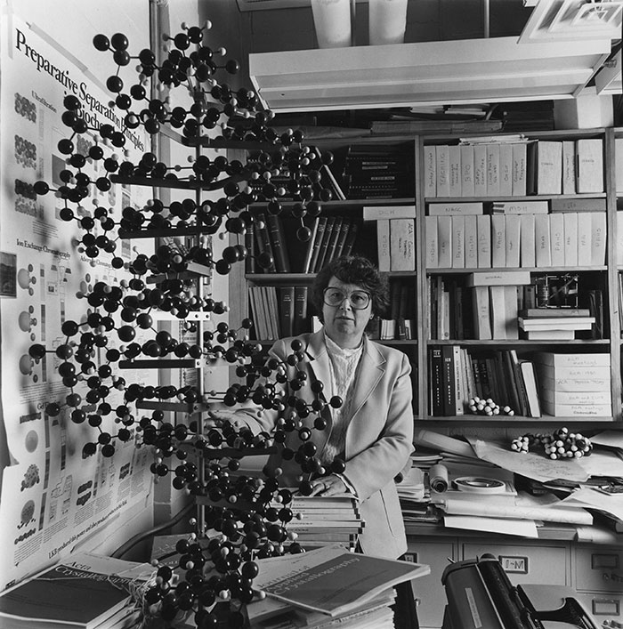
Jenny P. Glusker in her laboratory at the Institute for Cancer Research (Philadelphia) in 1980 or after. "The model was built by Ann Geale Diamond, Bob Diamond's wife, who lived in the next street to me in England and we went through the same schools and college. She was working for a model-building company in Cambridge, England and I only found out later that she had made it."
Some of the compounds involved in cancer, both chemical carcinogens and antitumor agents, captured our attention. Structure determinations for antitumor agents had started in Lindo Patterson's lab when Max Taylor, Eric Gabe, Jean Minkin and I worked on the structure of the copper-utilizing antitumor drug 2-keto-3-ethoxybutyraldehyde-bis(thiosemicarbazone). Later, the structure of estramustine, used to treat prostate cancer, was determined with Ken Tew at ICR, Beryl Hartley-Asp in Sweden who provided the material, and Bill Duax and the Buffalo crystallography group (who were working on the same compound). This study gave information on the conformation of this antitumor agent, how it might bind to its interaction target, and explained its chemical activity. A major class of compounds that we studied were the "ICR compounds" that had been devised at ICR by Hugh Creech, Richard Peck and Robert Preston of the chemotherapy group. These are acridine derivatives some of which showed antitumor and/or mutagenic activity. The antitumor agent ledakrin that we studied in collaboration with its originator, Professor Andrzej Ledochowski, from Gdansk, Poland, was a member of this class of compound. ICR compounds were presumed to interact with DNA, intercalating, as does acridine, between the bases of DNA; the side chains of these agents could then provide further action, such as alkylation of DNA. We determined the conformations and extents of overlap of their ring systems in packing in the crystalline state for several of these compounds.
We also tackled chemical carcinogens, starting with polycyclic aromatic hydrocarbons (PAHs) such as benzo[a]pyrene and 7,12-dimethylbenz[a]anthracene (DMBA). Such carcinogenic PAHs are oxidized in the body and the active agent is an alkylating agent such as a diol epoxide. We wanted to see if we could contribute to an understanding of their reactions in the body. Several PAHs become more carcinogenic when they are methylated at certain positions in the chemical formula. Such methylation may make it impossible, for steric reasons, for the substituted PAH molecule to remain totally planar. We wondered whether the twisting that results from this methylation enhances their possibility of interaction in the helical structure of DNA and what the effect was on the charge distribution in the molecule, that is, the ability of a metabolite to act as an alkylating agent, which is believed to be its function in cancer. Crystal structures of several metabolic products of carcinogenic PAHs that we determined included a diol epoxide of 5-methylchrysene, a presumed activated metabolite of a PAH more carcinogenic than chrysene itself. This work was carried out in collaboration with Maurice Coombs (Imperial Cancer Research Fund, London, England), Anthony Dipple and Robert Moschel (NCI-Frederick Cancer Research and Development Center), Ronald Harvey (University of Chicago) and Mahesh Lakshman (The City College and The City University of New York) and was done by Bud Carrell, Dave Zacharias, Setsuo Kashino (who spent a year at ICR as a Visiting Scientist from the University of Okayama, Japan), Maria Flocco, Kay Obendorf and Carol Afshar. A detailed electron-density study of DMBA with Cheryl Klein and Ed Stevens resulted from high-resolution X-ray data collection and multipole refinement of the structure. Assuming that the action of a carcinogenic PAH involves interaction of a PAH metabolite (presumably formed by an interaction with cytochrome P-450), we synthesized and studied the structures of some adducts of PAHs with nucleoside portions of DNA. We measured the extent to which the PAH ring system lay between the bases of the nucleoside. This was an extensive study carried out in collaboration with John Stezowski at the University of Stuttgart, Germany and later at the University of Nebraska in Lincoln.
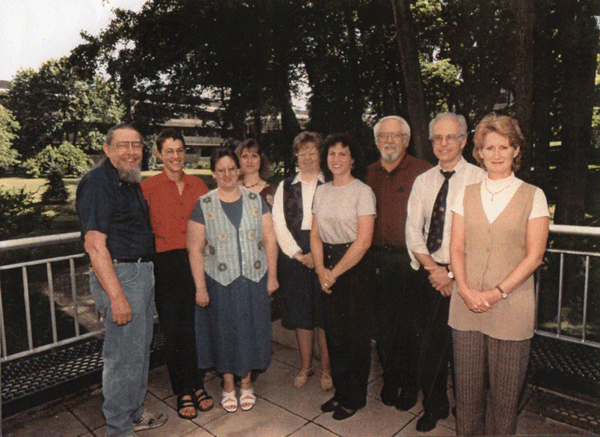
The Glusker lab group in 1990. Dave Zacharias, Trixie Wagner, Carol Afsher, Liat Shimoni Livny,
This led to a general interest in intermolecular interactions since structural studies reveal not only what a molecule looks like, but also how it interacts with other molecules (generally in a crystal of the same kind). Our investigations of these were greatly helped when Peter Murray-Rust came to our lab on a sabbatical. We investigated the three-dimensional geometry of the manner by which oxygen and nitrogen atoms in molecules bind to atoms in other molecules. We represented the results of our analyses in probability plots (looking like electron-density maps). Such analyses required the use of crystallographic databases, such as the Cambridge Crystallographic Data Base in England and the Protein Data Bank in the U.S.A. We then examined, in a similar manner, several other types of intermolecular interactions, including the locations of metal ions around carboxylate ions and near heterocyclic ring systems. Each relevant published crystal structure was examined in detail to ensure that we knew the coordination number of the metal ion and its geometry; results of such investigations were provided to the appropriate database staff. Results were then used in theoretical density functional calculations in order to obtain the energies of various states. Of particular interest to date were the surroundings of divalent Mg, Mn, Ca, Zn, and Pb (which has an interesting lone pair of electrons), and trivalent aluminum. Studies of monovalent sodium are in progress. We were able to show how different Zn2+ and Mg2+ are when they bind ligands such as water. The binding capacities of metal ions in various ionization states and coordination numbers were represented in triangular plots with oxygen, nitrogen and sulfur at the three corners of the triangle. They showed where the metal ion lay with respect to these three most likely binding atoms in proteins. These plots serve as useful signatures of each metal ion in each of its possible valence states. For example, Mg2+ binds the oxygen atoms of six water molecules in an octahedral arrangement, whereas Zn2+ can have a coordination number of 4, 5 or 6 and will bind nitrogen or sulphur as well as oxygen. Theoretical studies showed that divalent and trivalent cations will bind water molecules with their hydrogen atoms pointing away from the positive charge of the metal ion. Monovalent cations do not have the full extent of this power, while quadrivalent cations may tend to initiate a chemical reaction between water and the metal ion. This suggests why so many enzymes utilize divalent metal ions to bind their substrates and, often, to aid in the catalytic mechanism. These studies were done by Amy Katz, Bud, Christopher and Andrew Carrell, Jonah Erlebacher, and Liat Shimoni in my lab, Philip George at the University of Pennsylvania, Charles Bock at Philadelphia University and Gautam Desiraju (Visiting Scientist from the University of Hyderabad, India) and Colin Kennard (on sabbatical from the University of Queensland, Brisbane, Australia).
The enzyme structure that we have been studying through the years is D-xylose isomerase which converts D-xylose to D-xylulose and D-glucose to D-fructose. It is used industrially for the conversion of glucose to fructose to obtain high-fructose corn syrup for soft drinks and so is also referred to as "glucose isomerase." Albert Mildvan, an NMR spectroscopist at ICR (now Fox Chase Cancer Center), found crystals of this enzyme in the tube he was using to measure NMR spectra and he gave them to us. Work on this enzyme through the years at ICR has involved Bud Carrell, Helen Berman, Byron Rubin, Teresa Hurley, Trixie Wagner, Henry Katz and Helga Hoier, and collaborations with Jean-Francois Biellman (then at the Université Louis Pasteur, Strasbourg, France) and Carl Batt (Cornell University). The mechanism of action of D-xylose isomerase involves binding of the sugar substrate, opening of the sugar ring system, isomerization of the sugar and possibly cyclization, and then ejection of the sugar from the active site. In the X-ray structure of the enzyme at 0.94 Å resolution it was found that a truncated substrate had been bound to the metal ion, but that this ligand lacked atoms in the isomerization region of a substrate. As a result enzyme side chains in that region were disordered in the high-resolution map and this gave some hints as to how they might move during the catalytic process. Time-of-flight neutron diffraction studies of this enzyme, in collaboration with Gerald Bunick, Benno Schoenborn, Paul Langan and Andrej Kovalevsky at Los Alamos National Laboratory, showed the ionization states of the various side chains of the enzyme; of particular interest are lysine, histidine and carboxylic acid groups. In one structure a deprotonated water (that is, a hydroxyl group) is located near the site of sugar isomerization, suggesting how a hydrogen atom might be transferred from one carbon of the substrate to its neighbor. By varying the metal ions and ligand it was found to be possible to view various stages of the catalytic mechanism. So, if by chance one can obtain large enough crystals, the combination of neutron and high-resolution X-ray studies will contribute greatly to the elucidation of an enzyme mechanism.
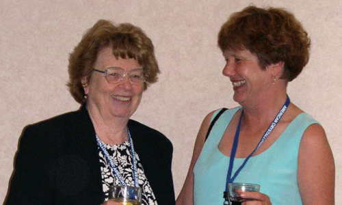
Jenny Glusker and Penny Codding, 2003. Each served as President of the ACA, Jenny in 1979 and Penny in 1998.
I have greatly enjoyed my career as a crystallographer and a member of the American Crystallographic Association and thank all of you for the honors and tasks that have been bestowed on me through the years. As asked, I can list a few of the tasks I have carried out, such as Newsletter editor, representative to the American Institute of Physics, Program Chairman of the Penn State meeting, President in 1979, Secretary Treasurer and Chairman of the U.S. National Committee for Crystallography and Editor of Acta Crystallographica D (macromolecules). There were two ACA meetings in 1979, one in Hawaii and one in Boston. It was a pleasure to finally be able to lure some Chinese crystallographers, especially Professor You Chi Tang who had studied years before at Caltech, to the Hawaii ACA meeting. When I stood up to introduce this ACA meeting the electric power to the entire university failed for a considerable period of time, delaying the start of the program. However Hawaiian dances at the banquet were appreciated by all. The Boston ACA meeting was exciting because so many protein crystallographers came and organized a wonderful program. Many of them had not been to an ACA meeting for a while and we were glad to welcome them back. I also, when vice-president, had to arrange the Alabama meeting and shortly before it was due to take place the main hotel in the location that had been planned for the meeting was destroyed in a storm and we had to move the meeting inland to Eufaula, Alabama (1980). This was a different but delightful location. As Chairman of the U.S. delegation to the IUCr meeting in Hamburg, Germany in 1984 I organized a party in the U.S. Embassy for members of all the delegations of other countries that attended the Congress, which served to bring these delegates together in a social event. I have been interested in the teaching of crystallography and served as Chairman of the Teaching Commission of IUCr for several years and organized schools in Egypt, Thailand, the Chinese University of Hong Kong, Tianjin (China), Madras (India) and recently spoke at a crystallographic meeting in Turkey. Ken Trueblood and I wrote a text on X-ray diffraction of crystals and it was expanded in a later text with Miriam Rossi and Mitch Lewis. Finally, I will still be teaching this summer (2011) at the ACA Summer School for Small Molecules run by Charles Lake and Bryan Craven.
—Jenny P. Glusker, 2011
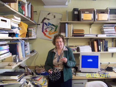
Jenny P. Glusker in her office at the Institute for Cancer Research, Fox Chase Cancer Center, Philadelphia, in 2011.
1]ICR = The Institute for Cancer Research in Fox Chase, Pennsylvania, now part of Fox Chase Cancer Center. |